Leiden researchers can now visualise the connections between brain cells. They do so using a microscopy technique in which Leiden physicists excel. This breakthrough could significantly advance the human quest to understand brain functions.
How does information flow through the brain? To understand this, researchers map the brain at every scale, from small networks of cells to the entire nervous system. This provides insight into how our brains work and how connections between cells may become disrupted in disease.
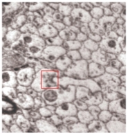
The research group led by Professor Sense Jan van der Molen uses a microscope that reveals how a brain structure is built. It can do so down to the level of a synapse, the tiny junction through which one neuron communicates with another cell.
Introducing the PEEM microscope in brain research
In a joint Chicago-Leiden project, Photoemission Electron Microscopy (PEEM) is introduced as a new tool for imaging brain tissue. In PEEM, the photo-electric effect is used to create images of materials of choice. Now, the researchers have applied it to image ultra-thin brain slices, taken consecutively from the same mouse brain.
As a result, high-resolution images were produced, at a faster rate and lower cost than mainstream imaging methods like transmission electron microscopy (TEM) or scanning electron microscopy (SEM). Once a consecutive set of thin slices is taken, a 3D model of this part of a brain can be reconstructed.
Tuesday Talk - Microscopy reinvented: peeking into living worlds
On the 9th of December 2025 professor Sense Jan van der Molen gives a Tuesday Talk at the Faculty of Science on his research.
Offering faster and cheaper 3D brain imaging
The ambitious effort to chart every neuronal connection in the brain - connectomics -- has long been held back by slow, expensive, and technically demanding imaging workflows. In this proof-of-concept study, PEEM truly changes the equation. Compared to traditional electron microscopy, it lowers both cost and technical barriers.
'At the current resolution of the images - 20 nanometres - synapses can already be identified. We managed to visualise these junctions where electrical or chemical signals pass from one neuron to another', explains Van der Molen. 'PEEM delivers fine structural detail of the brain without the heavy staining and complex sample preparation required by traditional methods. This new method opens the door to future high-throughput brain mapping.'
Optimizing resolution and samples
Another positive outcome is that PEEM images can be further improved in the future - both in terms of resolution and by preparing samples in ways that are best suited for the PEEM technique. In the coming years, the team will continue its collaboration with researchers from Chicago. Together with new Leiden postdoc Simona Borrelli, the team will produce images using a next-generation PEEM microscope, allowing for even sharper and more accurate images.
A catalyst for multidisciplinary discoveries
The Van der Molen Lab are building a strong reputation for integrating advanced microscopy with cutting-edge biology and biomedical research. They see enormous potential in combining PEEM imaging with research in biomedicine and beyond.
Proceedings of the National Academy of Sciences
Photoemission electron microscopy for connectomics
Auteurs: Gregg Wildenberg, Kevin M. Boergens, Amin Moradi, Rudolf Tromp, Sense Jan van der Molen, et al.


