Researchers at the University of Exeter and the Vanderbilt University Medical Center have collaborated to make a collection of images of pancreatic tissue from individuals with recent-onset Type 1 diabetes available in Pancreatlas, the world's first online imaging database of human pancreatic tissue created and housed at VUMC.
The most extensive image bank of samples of the pancreas from children who developed diabetes shortly before death has gone live at Vanderbilt University Medical Center, with the aim to advance global medical research in the diabetes field.
The University of Exeter and Vanderbilt University Medical Center (VUMC) teamed up to make high-resolution images of pancreatic tissue available in Pancreatlas, the world's first on-line imaging database of human pancreatic tissue created and housed at VUMC.
The pancreas contains the beta cells that produce the insulin that controls blood sugar and because of this is implicated in diseases such as diabetes. The image bank is a valuable asset to researchers because the human pancreas cannot be safely biopsied, and study of the cellular changes that cause type 1 diabetes can only be undertaken in pancreas specimens from individuals with the disease following their death.
The Exeter Archival Diabetes Biobank (EADB) includes 345 images of post-mortem tissue procured by former University of Glasgow professor Alan Foulis in the 1980s. These images represent the largest collection of pancreas samples recovered from patients who died shortly after the diagnosis of type 1 diabetes.
Foulis bequeathed the tissue collection to the University of Exeter Medical School upon his retirement, and the specimens were then digitized by Professor Peter In't Veld of Vrije Universiteit Brussel. The collection's curators, Exeter professors Noel Morgan, PhD, and Sarah Richardson, PhD, contacted the Pancreatlas creators to see if the images could be added to the online database and made available to anyone in the world. The Exeter professors then facilitated the image transfer.
"We are thrilled that this invaluable archive is now accessible to investigators throughout the world via the Pancreatlas platform," said Diane Saunders, PhD, research assistant professor of Medicine and co-scientific director of Pancreatlas. "Not only is this a unique group of samples, but the Exeter Archival Diabetes Biobank has enabled foundational advancements in human pancreatic research, including the profile of insulitis and evidence of viral involvement in driving beta-cell autoimmunity at the islet level. It is our expectation that these images will continue to spur scientific discoveries as investigators work to demystify the development of type 1 diabetes."
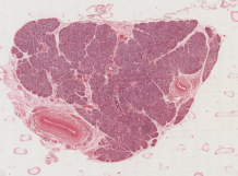
The EADB's 345 images represent 189 different donors ranging in age from three month up to 17 years. The onset of type 1 diabetes ranges from within days of their death to 19 years' duration. The images are of tissue sections stained using hematoxylin and eosin (H&E) or by immunohistochemistry targeting protein biomarkers to hormones and other signaling proteins.
"Mercifully, most people do not die until long after their diagnosis of type 1 diabetes, but this means that the availability of organs in which the disease process is still active is very rare,'' said Morgan, professor of Endocrine Pharmacology at the University of Exeter. "As a result, very few people across the world who are studying type 1 diabetes have been able to study tissue from young children with recent-onset type 1 diabetes.
"The provision of these unique EADB records via Pancreatlas is a major development because it provides easy access to a global audience and sets this collection in the context of other extremely important pancreas image collections available on the site. The search criteria and annotation systems developed by the Vanderbilt team enable researchers to select the images most relevant to their own studies and to view these in high resolution from their own desks."
In total, Pancreatlas currently hosts 2,180 images, representing several biobanks and research initiatives. The site provides the scientific community access to annotated images of human pancreas tissue and associated donor characteristics with the hope of advancing the understanding of diseases such as diabetes, pancreatic cancer and pancreatitis.


