EMBL's imaging technology helps researchers gain insights in the fungus' journey from the lung to the brain
Found all over the world, the fungi cryptococcus usually doesn't affect healthy individuals. However, it sometimes causes severe meningitis in immunocompromised individuals, like people with HIV. Infection usually occurs when people inhale the fungi, spreading it to the lungs and brain, possibly with fatal consequences.
Greetje Vande Velde, Research Professor at KU Leuven, has studied cryptococcus for almost a decade. How the fungi are able to cross the blood brain barrier is still unknown, and that's why Vande Velde and Jim Swoger, Head of the Mesoscopic Imaging Facility at EMBL Barcelona, started collaborating several years ago.
In 2015, Vande Velde was a postdoc at KU Leuven and Swoger still worked in the Sharpe Lab, at the Centre for Genomic Regulation (CRG) in Barcelona. She spent a research fellowship in the Sharpe lab to visualise mouse organs in 3D with Swoger, using the recently invented techniques Optical Projection Tomography (OPT) and Selective Plane Illumination Microscopy (SPIM).
"Greetje's expertise is in 3D in vivo imaging, which allows for live observation of disease spread, but it doesn't allow us to look at cells themselves within the context of organs and tissues," Swoger said. "OPT and SPIM allow us to image complete mouse organs ex vivo at high resolution in 3D and help us see the spatial organisation of the cryptococci."
In 2017, EMBL Barcelona and the Mesoscopic Imaging Facility (MIF) were established, with Jim Swoger as Head of MIF. The project then went a step further by using a combination of the two imaging technologies used at the CRG: OPT plus SPIM. This became the hybrid OPTiSPIM. SPIM provided high resolution, which complemented the imaging speed and multi-modality (fluorescent and absorbing contrasts) of OPT.
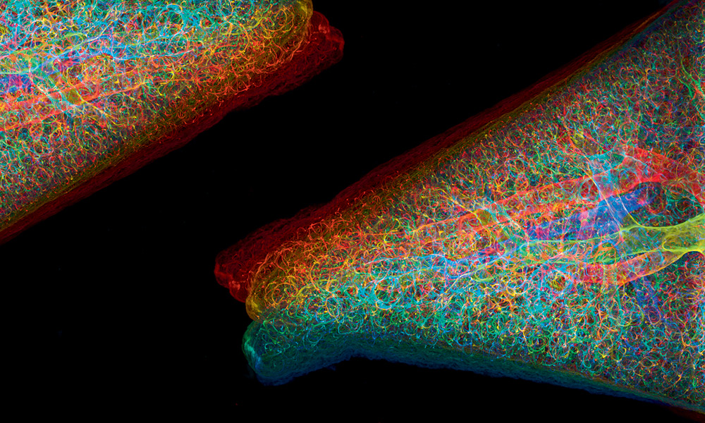
Vande Velde and Swoger imaged entire mouse lungs and brains infected with cryptococci to get 3D images at cellular resolution. They could then observe the fungi in the context of entire organs and see how fungal populations were organised in the folds of the brain.
These imaging technologies and the sample preparation protocols allowed the researchers to make novel observations on where and how the fungi assembled - something that hadn't been captured previously. Cryptococcus populations were generally organised around the microvasculature of the brain, which is the system of tiny blood vessels like capillaries and venules. Such observations are pivotal to further probing how the fungi enter and infect the brain.
"The collaboration with the MIF helped us achieve important insights towards understanding the disease pathogenesis." Vande Velde said. "There have been cases of apparently healthy individuals with severe cryptococcal infections that challenge our understanding of this disease to this day. We need to tackle this and other infectious diseases without delay."