Benign and cancerous calcium phosphate deposits that may look identical on a mammogram have distinct differences in their structures and formation processes, researchers at the University of Illinois Urbana-Champaign and collaborators at the Mayo Clinic and the University of Texas at Austin found in a new study that provides the first detailed descriptions of how calcifications form in breast tissue. The findings suggest new diagnostic criteria that could lead to fewer benign biopsies and guide therapeutic development, the researchers say.
"Dense calcifications are very common in breast tissue. They are seen easily on a mammogram, which doctors can use to classify benign, probably benign and suspicious categories," said study leader Bruce Fouke, a U. of I. professor of earth science and environmental change and director of the Roy J. Carver Biotechnology Center at Illinois. "But most biopsies of spots deemed suspicious end up being benign, meaning those patients underwent painful procedures unnecessarily. We want mammograms to be more precise and more accurate for distinguishing between benign breast disease and cancer."
Fouke's group has pioneered the field of "GeoBioMed," a combination of geology, biology and medicine, and previously applied it to the study of kidney stones and calcifications in the heart.
The new study examined biopsied tissue samples of benign breast disease and ductal carcinoma in situ that had been removed when patients underwent surgery as part of a long-term Mayo Clinic study. To document the mineral characteristics of BBD and DCIS, the researchers used 12 different methods to characterize the samples, including a suite of light, laser and electron microscopes and X-ray and Raman spectroscopy techniques.
"We have developed this analytical arsenal for understanding complex mineralization pathways," said Mayandi Sivaguru, the first author of the paper and the director of the Cytometry and Microscopy to Omics Facility in the Carver Biotechnology Center. "Previous research has often used only a couple of standard techniques, which often miss a comprehensive contextual evaluation of breast health and disease progression. We needed a holistic approach to see the whole picture of BBD and DCIS development that is recorded inside calcifications and the detailed history of how they formed."
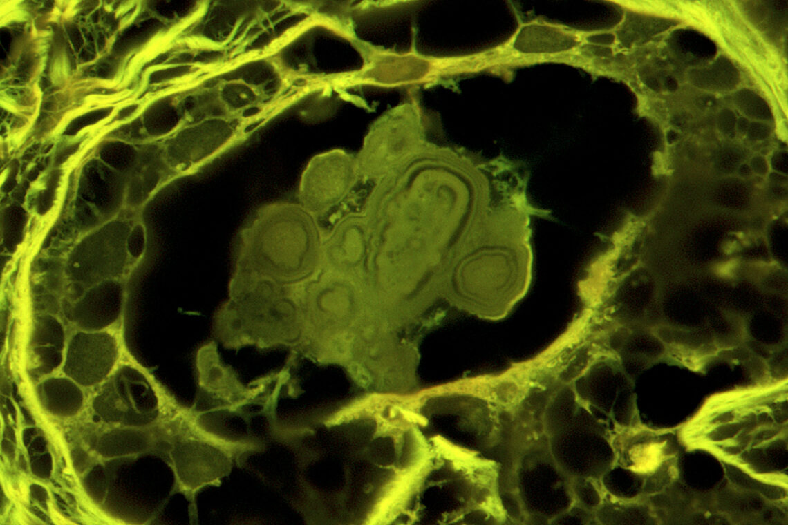
The researchers found that the calcifications were made of amorphous calcium phosphate, a mineral with the ability to shapeshift and rearrange, though it had long been assumed to be the crystalline calcium phosphate type found in bone, hydroxyapatite. The team analyzed the layers of the ACP deposits to trace how they began as small spherules that coalesced into nodules. The nodules then entombed cells and incorporated other molecules, such as proteins, waxy substances and cholesterol.
The shape and progression of calcification differed in BBD and DCIS samples. For example, BBD had more spherical nodules with concentric layering, while cancerous calcifications tended to be more elongated and irregular. Some cancerous nodules also showed progression similar to fossilization characteristic of petrified wood, Fouke said.
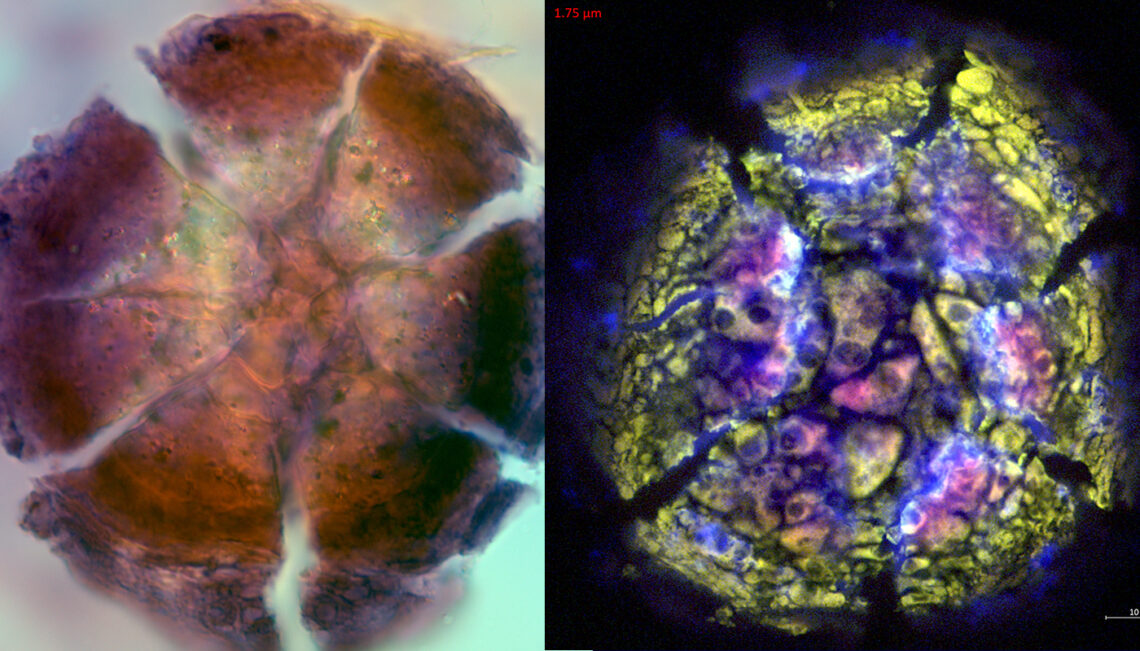
"The types of ACP nodules we saw were completely unknown and establish a brand-new classification scheme between BBD and DCIS," Fouke said. "Each has a different genesis and history of formation, reflecting changes in breast physiology that in turn correlated strongly with whether a biopsy sample was designated benign, possibly benign or suspicious."
Knowing that the calcifications are made of ACP rather than crystalline hydroxyapatite opens the possibility of treating BBD, as certain drugs are known to dissolve ACP deposits. If benign calcifications could be dissolved, then misidentifications in mammograms would be rarer and millions of unwanted biopsies could be prevented, Sivaguru said.
"The collaboration between our teams brought modern analytical tools to link geology knowledge and public health," said study co-author Rohit Bhargava, the director of the Cancer Center at Illinois and a professor of bioengineering. "The unique collaborations at our university and the great partnership with Mayo Clinic researchers has the potential to lead to better breast cancer care by understanding cancer from a unique perspective."
Next, the researchers will work to characterize calcifications in more advanced invasive breast cancer and to document whether and how calcification plays a role in DCIS progressing to invasive breast cancer. They also hope to study ACP calcifications with a GeoBioCell, an experimental microfluidic device the Illinois team developed.
"It's a roadmap for the future of controlled experimental testing. For instance, if we want to know, if a woman was to drink more water, would it make a difference in the amount or type of breast calcifications? We could flow more or less water and breast fluids through samples in the GeoBioCell and track how the calcifications grow. Similarly, we could test the effectiveness of drugs and combinations of different plant extracts to find new therapeutic agents," Fouke said. "Our aim is to predict and ultimately prevent breast calcifications, reduce inaccurate mammogram diagnoses and lay a framework for therapy development."
The Barbara and Ed Weil Foundation supported this work. Fouke is the Ralph E. Grim professor at Illinois and a member of the Cancer Center at Illinois.


