Scientists have long used fluorescent dyes—which emit light—to research cell structures and activities. By attaching fluorescent molecules to a protein, for example, scientists can take images of it under a microscope to track its movements and study its functions.
The problem with conventional fluorescence imaging, however, is that it's impossible to distinguish between structural features that are separated by distances shorter than half of the wavelength of the emitted light. Similar to the way two oncoming car headlights at a certain distance appear to merge into one, two structures close together within a cell that are both emitting light will appear indistinguishable.
"Conventional fluorescence imaging is missing a lot of information because of that," explained Françisco Raymo, a professor in the Department of Chemistry at the University of Miami College of Arts and Sciences.
Fortunately, Raymo and his team in the college's Laboratory for Molecular Photonics have developed a solution to this problem.
They have designed a family of molecules that are photoswitchable, meaning that they can be switched on and off using light. The molecules glow when they are exposed to both green and yellow light and return to their non-luminous state upon further exposure to yellow light. This enables researchers to switch the molecules on and off independently so that only one is visible at a time, allowing for fluorescence imaging that can distinguish between individual molecules and provide an incredibly detailed image.
"You can attach these molecules to proteins, to DNA, to the membrane of cells, to many different targets at the same time, and then you can, by switching them on and off, reconstruct images with resolution at the nanometer level," Raymo explained. "Using the same idea, you can watch them move in space, so you can track their dynamics. You can use this to study how biomolecules behave inside a cell, with a resolution that is impossible to achieve with conventional fluorescence imaging."
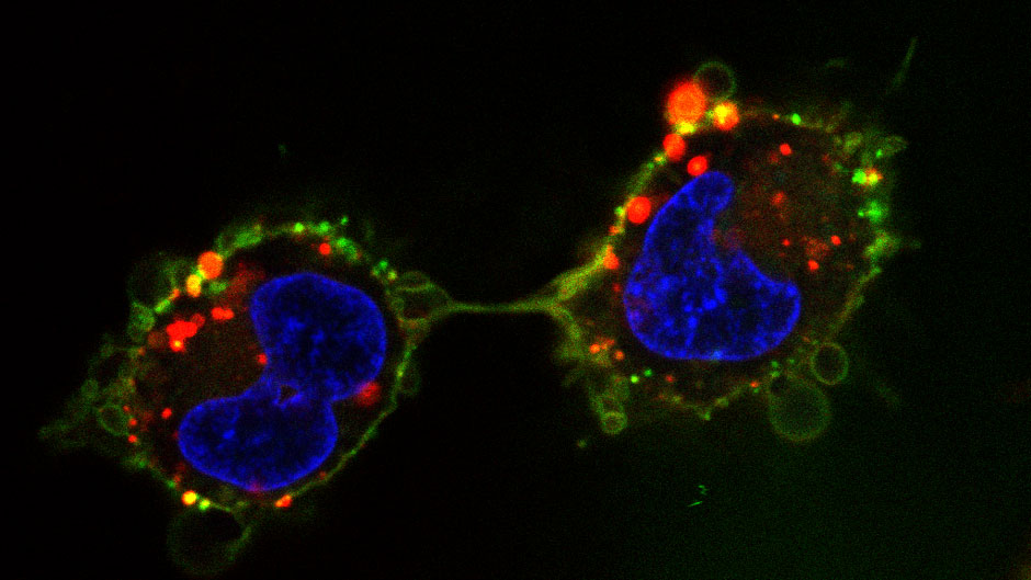
Human osteosarcoma cells labeled with fluorescent dyes, including one of Raymo's photoswitchable dyes, which glows green to illuminate the plasma membrane. Photo: University of Miami Laboratory for Molecular Photonics
The potential applications for these molecules include using them to image diseased cells to better understand their behavior and how it differs from that of normal cells.
Raymo is currently part of an interdisciplinary consortium based at Northwestern University that is using the molecules to study cancer stem cells with the goal of developing new treatment strategies.
Although Raymo acquired an interest in chemistry at a young age, he didn't set out to design molecules with biomedical applications. His initial interest was in developing molecules that could be used for computing.
Raymo grew up in Messina, Italy, where he often spent time in the laboratory of his grandfather, an organic chemistry professor at the local university.
"I kind of grew up in a chemistry lab," he said.
After completing an undergraduate degree in chemistry at the University of Messina and a Ph.D. and postdoctoral fellowship at the University of Birmingham in the United Kingdom, Raymo moved to the United States for a postdoctoral fellowship at the University of California, Los Angeles. He arrived at the University of Miami in 2000, where his research initially focused on developing photoswitchable molecules for computing.
Then, in 2006, Raymo read an article that changed the focus of his research. The article was about photoswitchable proteins that behaved similarly to Raymo's molecules and could be used for biomedical imaging.
"I realized that our compounds can be used for the same applications with advantages that only synthetic dyes can offer," Raymo said. "So even though I was using the same molecules, I moved my long-term goal from computers to biomedical applications."
Raymo adjusted his molecules, which were originally developed with funding from the National Science Foundation, to tailor them for biomedical imaging with support from the National Institutes of Health.
It was a prescient shift. The scientists who developed the photoswitchable proteins that Raymo had read about in 2006 went on to win the 2014 Nobel Prize in Chemistry. The field of super-resolution imaging has since "exploded," Raymo said, with chemists working to make similar molecules, engineers designing new microscopes, and biologists applying these cutting-edge tools to their work.
The applications for these molecules aren't limited to biology. Raymo has also developed photoswitchable fluorescent molecules for other applications. In collaboration with Peter Minnett, a professor emeritus at the Rosenstiel School of Marine, Atmospheric, and Earth Science, Raymo designed and used molecules to measure temperature at the micrometer level—what he described as something akin to "molecular thermometers"—with support from a National Aeronautics and Space Administration (NASA) grant. He currently has a grant from the Army Research Office to use photoswitchable molecules to develop micro-lasers.


