New findings illuminate mechanisms of protein production, transport, and folding in bacterial cells, but also establish a new way to study cellular 'cities', exploring molecular 'communities' rather than molecules in isolation
Summary
- Two recent publications from EMBL Heidelberg's Mahamid Group have improved our mechanistic understanding of protein production and transport across the membrane in a minimal bacterium and visualised the complex interactions between molecular processes.
- Both studies lean into the increasing body of research that explores macromolecular systems in the larger context of intact cells, rather than in isolation.
- The researchers studied the 'factories' in cells that read genetic instructions and build proteins, revealing extensive cross-talk between these processes.
- They also investigated protein transport across the membrane and beyond the cell, discovering and characterising a previously unknown protein-folding machinery at the cell surface.
- Together, the research illuminates how different cellular processes interconnect and serves as a proof of concept for future studies in more complex cells.
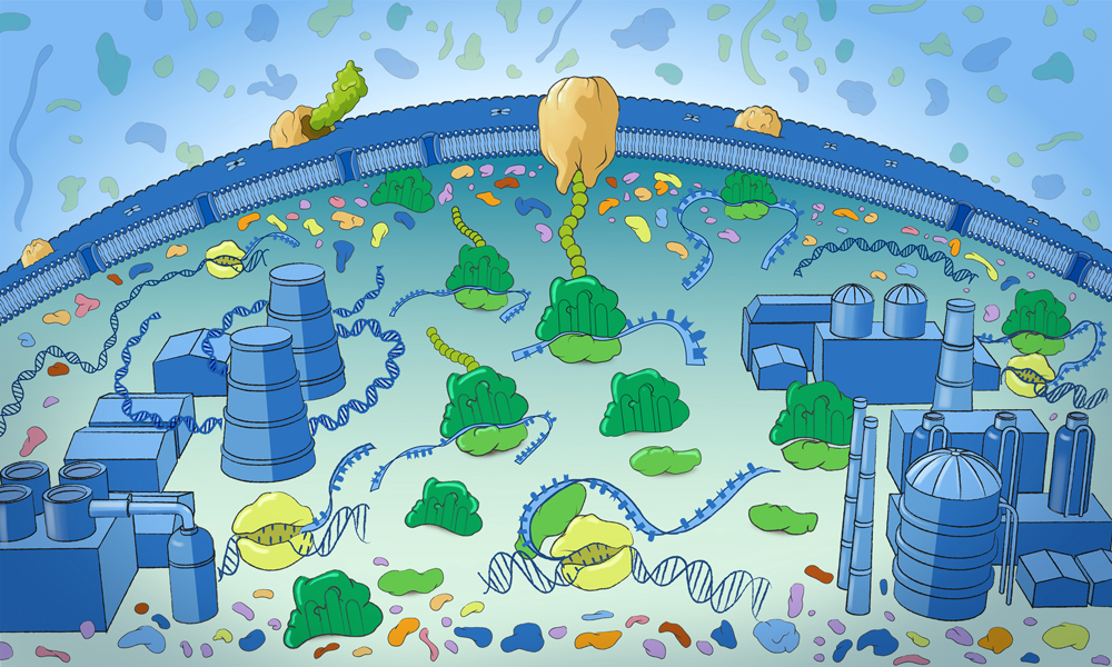
Scientists have often thought of cells as small bustling cities with parts that resemble factories, power grids, and shipping systems. And while past research has illuminated what we know about the various individual components of these cell cities, it hasn't had the tools to probe more deeply into how those different elements interact.
Researchers from Julia Mahamid's EMBL Heidelberg research group, however, have tapped the power of cryo-electron tomography (cryo-ET) to do exactly this: visualise how different cellular processes are connected. Their latest findings are published in two papers in the journal Cell.
"Both projects are essentially proofs of concept for this kind of analysis," said Joe Dobbs, lead author on one of the two papers and a former PhD fellow in Mahamid's group. He is currently working as a postdoctoral researcher at the Max Planck Institute for Brain Research in Frankfurt. "Rather than looking at a single type of molecule in isolation, both papers examine interactions between different cellular systems with a common thread of understanding how different processes are coordinated."
Getting to know protein production better
Dobbs and colleagues focused on imaging Mycoplasma pneumoniae bacteria using cryo-ET. Cryo-ET is a powerful imaging technique that allows scientists to see inside flash-frozen cells in 3D and in great detail, revealing structures of molecular machines.
The scientists used this technology to reconstruct high-resolution maps and count instances of the protein production machinery (ribosomes) in different functional states across hundreds of individual cells. Because proteins do much of what a cell needs to stay alive, understanding this process helps us learn how cells work, why diseases happen, and how to develop better medicines.
Among other findings, the scientists observed several new complexes that directly connect transcription - the process of producing mRNA from DNA - with translation, the process of producing proteins. This allowed them to better understand how protein production is regulated, to provide structural evidence for long-hypothesised 'supercomplexes', and to suggest new molecular mechanisms for how the intertwined processes are controlled.
Additionally, they found parts of the ribosome attached to the cell membrane even when they weren't producing proteins. Their results suggest that these subunits might detach from the membrane only when conditions allow a new round of protein production to begin. This behaviour is similar to what had been observed in mammalian cells decades ago, and the new findings suggest that such mechanisms may be conserved from bacteria to humans.
"I think of it as a city," Dobbs said. "The molecular machines I work on are like little factories that produce things, while some of my colleagues have been studying systems that transport and process those products. Together, they form different parts of a living molecular community inside the cell - much like a city with different specialised activities all working together."
This focus on the interaction between the molecular machines, rather than in isolation, is part of what makes the two papers novel.
Discovery of wholly new complexes at the cell's border
Elsewhere in the Mahamid group, Rasmus Jensen was leading research on a system responsible for protein transportation and folding at the cell membrane. He wanted to better understand what happens to proteins after they're produced.
"Joe started with a biological question he wanted to answer," Jensen explained. "I started from the opposite direction. We'd seen something interesting in the cell and worked backwards to figure out what it was, eventually discovering that it's a new configuration of a molecular machine that helps proteins either be exported out of the cell or inserted into its membrane."
By combining cryo-ET imaging, proteomics, and computational protein structure predictions, the team found that this machinery contains the well-known Sec translocon, a protein channel in the cell membrane that acts like a doorway to move newly made proteins out of the cell, along with three previously unknown proteins that help these transported proteins fold into their correct functional shapes.
Cryo-ET allowed the researchers to create the most detailed images so far of the conserved part of the bacterial protein transport machinery. These images showed how proteins translated by ribosomes begin moving through the cell membrane and revealed that protein transport is closely linked to a newly discovered protein-folding system outside the cell in Mycoplasma pneumoniae.
"Working together with many colleagues at EMBL, we combined expertise in microbiology, proteomics, bioinformatics, structural biology, and integrative modelling," said Mahamid, EMBL Group Leader and senior author on both papers. "This paper tells the entire story of the discovery process: visualising a new cellular machinery, identifying the genes and proteins, and figuring out how it all functions. That's a remarkable achievement and opens new ways to identify new complexes and functions in different systems."
From simple bacterial cells to more complex species
While the discoveries are interesting in their own right, these studies more importantly lay the conceptual foundation for future studies.
"In both Joe's work and mine, we've started with what we'd call a minimal cell where everything is relatively simple," Jensen said. "We've advanced what is conceptually possible in this minimal bacterium and established ways to apply these methods to more complex organisms."
"The future is studying molecular communities," Mahamid said. "Decades of structural biology done on isolated molecules provided us with detailed views of their structures and functions. In the past, we just didn't have the technology to study their more complex interactions and cross-talk. This work points again to how cryo-ET is an enabling technology, allowing us to see new things and connectivity inside cells. Its ability to show us more seems only limited by our asking the right questions."