Adapted from an article by Robert Sanders at UC Berkeley.
Nearly 100 years ago, a discovery revolutionized light microscopy. The introduction of the phase-contrast microscope, which garnered a Nobel Prize in 1953, brought into clear view structures inside cells that had previously been too faint or washed out for biologists to study.
A team of physicists from UC Berkeley and Lawrence Berkeley National Laboratory (Berkeley Lab), have now adapted the phase-contrast technique to cryo-electron microscopy (cryo-EM), which has about 10,000 times the magnification of light microscopy. As reported in Science, their laser-based phase plate produces sharp images of molecules that today's cutting-edge cryo-EM systems struggle to capture.
The new technology was brought to fruition by more than 15 years of theoretical and experimental work by leading microscopy scientists, collaboration with expert machinists, and support from Biohub. The phase plate is paired with a new, custom Thermo Fisher Scientific microscope that was developed to maximize the benefit of the plate's ultra-bright laser. Images taken by the system are notably clearer and sharper and contain greater detail that structure-solving software can process to generate more accurate atomic models of the molecules captured.
"Theia is the Formula 1 of microscopes. It has better resolution than the standard cryo-EM, even without the laser. With the addition of the laser phase plate, we hope that it really becomes one of the world's best instruments overall," said Holger Müller, a UC Berkeley professor of physics and faculty scientist in Berkeley Lab's Biosciences Area who led the development effort. "Before, studying structures with cryo-EM was like trying to look at paintings in a dark gallery. With Theia, it's like the lights have been turned on for the first time."
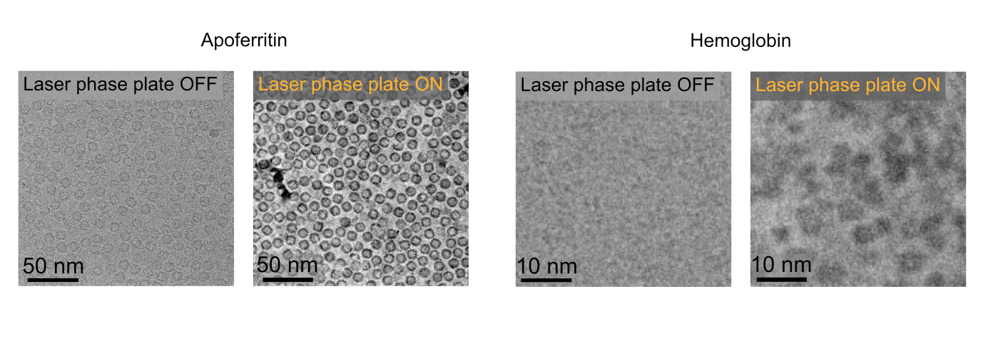
In their paper, Müller and his colleagues demonstrate the system's power by imaging aldolase, a protein in muscle that is relatively easy to capture with today's cryo-EM machines, and hemoglobin - a protein that carries oxygen in blood. Hemoglobin is a smaller protein that sits at the lower size limit for current machines and is often used as a benchmark for cryo-EM performance. The laser-phase plate improved the resolution of the protein structure in both cases, but more so for the hemoglobin. Their study also tested the microscope on samples of varying quality to demonstrate how the phase plate can alleviate the difficult and time-intensive process of sample preparation. Currently, single-particle analyses like these require thousands to millions of isolated target molecules that have been carefully frozen to -160 degrees C or below. And even the smallest amount of ice on samples can interfere with imaging.
"We had a whole spectrum from larger particles that are not that challenging with extremely good specimen preparation to challenging small particles with bad preparation. Of course, the better the specimen, the less important it is that you have a top microscope. It was the most challenging ones is where we see the strongest improvements," said Müller.
The system is currently installed at UC Berkeley, and the team is now working to expand the microscope beyond single-particle analysis to perform a newer technique referred to as cryo-electron tomography (cryo-ET). Similar to how CT (computed tomography) scans at the hospital generate images of body parts, cryo-ET assembles different angular views of a molecule or cellular structure into a 3D image. Cryo-ET will provide a huge leap in scientists' ability to study cellular processes because it captures molecules in their natural states inside cells, unlike single-particle cryo-EM, and offers much higher resolution than light microscopy.
"Single particle analysis and tomography offer different advantages in structural biology, and depending on the scientific question to be solved one or the other could be more suitable. In cryo-ET, the ability to look at cells in their native context is one of its main advantages," said Jessie Zhang, a postdoctoral researcher at UC Berkeley and in the Biosciences Area who is co-first author on the Science publication. "However cells can be very messy, and the low signal to noise in conventional EM makes interpreting tomograms a challenge. The laser phase plate has the potential to allow biologists to see and understand more of the proteome in action."
The evolution of biological imaging
Under a light microscope, animal and plant cells appear nearly transparent, yet it's difficult to get a good look inside. The tiny internal structures and organelles we want to study only scatter a small amount of light, making the view too low contrast to discern much detail. Chemical stains were developed to improve the contrast, but these additives also kill the cell.
In 1930, Dutch scientist Frits Zernicke realized that the brightness or amplitude of the light was not the only feature affected when passing through a cell. The scattered light is also slowed down, which shifts its phase - the timing of the peak of the waveform - by a small amount. While this phase shift is invisible to the human eye, it can be turned into visible contrast by also phase-shifting the non-scattered light by 90 degrees. When the scattered and non-scattered light are ultimately focused on the retina, features in the sample are enhanced relative to the background, boosting the contrast. Zernicke received the 1953 Nobel Prize in Physics for this discovery.
By the early 1940s the phase-contrast light microscope had proved its value. Scientists speculated about adapting this technique to the electron microscope, which uses a beam of electrons rather than a beam of light to image much smaller structures. The inherent contrast is very minimal in electron microscopy, so the scientific community was eager for solutions. But attempts to make a phase plate that shifts the phase of an electron beam reduced the beam intensity too much, made the images unstable, or resulted in lower resolution.
In 2010, Robert Glaeser, a long-time joint researcher who is now a professor emeritus of molecular and cell biology at UC Berkeley and retired senior scientist in the Biosciences Area, started a collaboration with Müller to develop a phase plate that avoided these pitfalls using an intense laser.
Glaeser is a pioneer of cryo-EM who has been advancing the approach since it was first invented in the 1960s. He helped overcome one of the initial challenges of cryo-EM - sample destruction by the intense electron beam - by designing a protocol of freezing samples to liquid nitrogen temperatures (-196 C, or -321 F). He also developed a method to combine thousands of images of frozen molecules to create highly detailed structures. The originators of cryo-EM were awarded a Nobel Prize in Chemistry in 2017, and both the Nobel Committee and recipients credited Glaeser's work.
After publishing a paper outlining the theoretical foundation of a laser phase plate in 2010, Müller and the team spent 16 years bringing it to life. With funding from the National Institutes of Health (NIH), his team tackled the first challenge: designing a continuous laser powerful and focused enough to shift the phase of an electron beam by 90 degrees. They achieved this, following ten years of research and development, by trapping a laser beam in a mirrored cavity that both focuses the beam and intensifies it as the light bounces back and forth more than 10,000 times.
"It's 75 kilowatts focused to a few microns," Müller said. "That's more power than a military laser. It builds up the brightest continuous laser focus ever."
They later proved that the entire laser phase plate works by installing it on the existing microscope in Glaeser's lab, another Thermo Fisher Scientific system that was state-of-the-art when their work began. But the phase plate's full potential could not be demonstrated without a custom microscope designed to pair with the technology's new capabilities. Around the time of these initial trials in 2018, biophysicists from across the Bay Area were brought together at a workshop hosted by the Biohub. The collected experts were thrilled by the team's progress and eager to see the technology fully realized. With funding from Biohub, the scientists acquired a Thermo Fisher Scientific microscope designed to accommodate the laser-phase plate. Müller named the complete system "Theia" after the ancient Greek Titaness of light and radiance.
Installation and final development were completed in 2025, with the help of resources from a Berkeley Lab Laboratory Directed Research and Development (LDRD) award. Within days, the scientists began producing striking high-contrast images. Soon after, they captured the series used in the proof-of-principle Science paper.
From Berkeley, onwards
Now, just over a year after the system first came together, the team is on the cusp of being able to use Theia to answer scientific questions. The Berkeley Lab and UC Berkeley scientists are currently working to maximize the prototype microscope's capabilities and improve its focus, which could double the amount of structural information in each image. The Biohub team members are developing technologies to integrate the laser phase plate for cryo-ET investigations.
And meanwhile, the scientists and machinists hope their collaboration with Thermo Fisher will result in streamlined versions of the system for other groups around the world.
"I like to say ours is not street legal," said Müller. "It's optimized for peak performance, so it requires a bit more user training. The other configurations of the microscope that are in the works are more user-friendly."
According to Müller, the unprecedented power of the original Theia is a testament to the level of expert design and craftsmanship that his colleagues put into the project over the years. "Our prototype was made by very skilled labor of very special human hands," he said.
This project was made possible by funding from the NIH, the National Science Foundation, the Gordon and Betty Moore Foundation, the Chan Zuckerberg Initiative DAF, and through and Berkeley Lab's LDRD program.
Cryo-EM is a technique for determining the structure of molecules that has been a major tool for biologists for decades, but truly began to revolutionize our understanding of proteins and accelerate new drug discovery starting in the 2010s, when advances led by Berkeley Lab scientist Peter Denes enabled atomic-level resolution for some samples. But to this day, it remains difficult to produce clear images of small molecules, such as human proteins. To avoid destroying the target molecules, a diffuse electron beam is used, but for molecules with fewer atoms, this produces small, faint scattering patterns ("signals") that are hard to distinguish from background noise.
Cryo-EM becomes very difficult for proteins around 70 kilodaltons, but around 90% of human proteins are smaller. To overcome this limitation, scientists must collect and painstakingly prepare thousands to millions of samples of their target molecule to statistically average the low-signal data in each image, and often need to use other molecules as physical scaffolding, which may distort the target molecule from its natural shape.
With the laser phase plate, it's now possible - though difficult - to image the benchmark molecule, hemoglobin. The team hope to improve their resolution to ~17 kilodaltons by pairing the phase plate with a focused electron beam. As counter-intuitive as it may sound, current cryo-EM systems use a defocused beam to generate contrast. But with the contrast provided by the phase plate, the system could be pulled into focus to deliver a two-fold boost in the signal-to-noise ratio.


