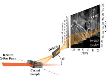
Dark-field X-ray microscopy views defects deep inside millimeter-thick crystals by capturing images of the X-ray diffracted beam.
A Lawrence Livermore National Laboratory (LLNL) scientist and collaborators have demonstrated the first ever "defect microscope" that can track how populations of defects deep inside macroscopic materials move collectively.
The research, appearing today in Science Advances, shows a classic example of a dislocation (line defect) boundary, then demonstrates how these same defects move exotically just at the edge of melting temperatures.
"This work presents a large step forward for materials science, physics and related fields, as it offers a unique new way to view the 'intermediate scales' that connect microscopic defects to the bulk properties they cause," said Leora Dresselhaus-Marais, a former Lawrence fellow and now assistant professor of Materials Science and Engineering at Stanford University.
Connecting a bulk material's microscopic defects to its macroscopic properties is an age-old problem in materials science. Long-range interactions between dislocations are known to play a key role in how materials deform or melt, but scientists have until now lacked the tools to connect these dynamics to the macroscopic properties.
Defects underlie many of the mechanical, thermal and electronic properties of materials. A prominent example is the dislocation, which is an extended linear defect in the atomic lattice that enables crystalline materials to permanently change their shape under loading. The range of hardness and workability in ductile materials occurs because of how their dislocations can move and interact.
In the new research, the team used time-resolved dark-field X-ray microscopy (DFXM) to directly visualize how dislocations move and interact over hundreds of micrometers deep inside bulk aluminum. With real-time movies, they showed that the thermally activated motion and interactions of dislocations that comprise a boundary and show how weakened binding forces destabilize the structure at 99 percent of the melting temperature.
The team resolved the individual and collective motion of the dislocations in a dislocation boundary (DB) beneath the surface of single-crystal aluminum. Their images map how the DB migrates along a very low-angle boundary as it is heated from 97 percent to 99 percent of the melting temperature (660 degrees Celsius). They then zoomed in on how dislocations enter and leave the boundary, causing two DB segments to coalesce and stabilize into one cohesive structure. As the DB subsequently migrates and increases its spacing between dislocations, they observed how the boundary destabilized.
"By visualizing and quantifying thermally activated dynamics that were previously limited to theory, we demonstrate a new class of bulk measurements that is now accessible with time-resolved DFXM, offering key opportunities across materials science," Dresselhaus-Marais said.
The team also includes scientists from Technical University of Denmark, Nevada National Security Site, CEA Grenoble, Universität für Bodenkultur Wien in Vienna and the European Synchrotron Radiation Facility. The work was funded by LLNL's Lawrence Fellowship and the Laboratory Directed Research and Development program.


