
We commonly say an art curator casts "an expert eye" over a painting, or an auditor over a set of accounts.
But what is it that, for example, allows an experienced birdwatcher to identify an uncommon species rapidly, at a distance, and against a chaotic background in the wild?
And what separates the gaze of a qualified brain surgeon from that of a first-year medical student? How is that kind of visual expertise acquired?
These questions are all highly relevant to the design and training of future artificial intelligence (AI) applications capable of analysing and interpreting complex medical images. Researchers in Macquarie University's Computational NeuroSurgery (CNS) Lab , led by Professor Antonio Di Ieva , have long argued the answers aren't just a matter of additional knowledge but also involve measurable, structural differences in how the eyes of an expert move and scan the relevant field for information.
Now they've been able to put hard data behind that idea with the publication of three new studies in which they harness the mathematical power of fractals to chart the development of visual expertise from novice to neurosurgeon.
Their work started with high-resolution tracking to record the eye movements of medical students, surgical trainees and qualified neurosurgeons as they viewed images ranging from simple X-rays to CT and MRI scans. Then they combined fractal mathematics and machine learning – a form of AI – to make sense of the overall pattern of those movements, translating the structure and complexity of visual scanning into geometric and numerical expressions.
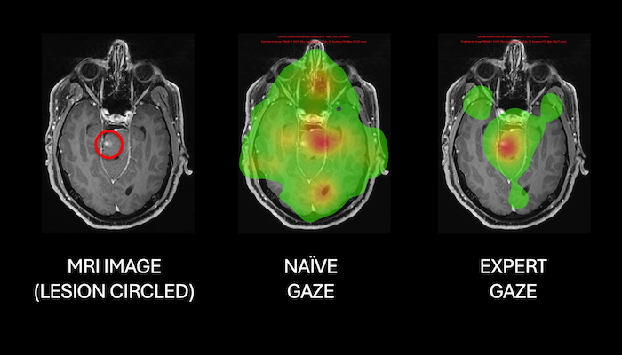
Heatmaps comparing eye gaze fixation between naïve and expert groups when looking at an MRI image showing a brain lesion. (Images supplied by Macquarie University CNS Lab)
"Fractal analysis is a mathematical way of measuring how complex and organised a pattern is," explains Dr Ghasem Azemi , Research Associate in the CNS Lab. "It goes beyond simply recording where or how long people look and helps reveal the extent to which their gaze is random or follows organised, repeating patterns."
In a study published in the journal Medical & Biological Engineering & Computing, CNS Lab researchers recorded the eye movements of 13 Doctor of Medicine (MD) students from Macquarie Medical School as they viewed a range of medical images and followed them for three consecutive sessions (semesters) of their training.
When combined into a single measure, the Fractal Eye-Gaze Expertise Index (FEI), the analysis showed a clear, statistically significant trend.
"As students progressed through the MD program, their gaze became less erratic and more structured, closer to the efficient scanning patterns of experts," says Dr Azemi. "This pattern suggests a shift from reliance on low-level image features to more targeted, diagnostic-driven visual processing. Fractal analysis allowed us to quantify that growing expertise."
A second study, published in the Journal of Eye Movement Research , helped broaden the lens further. PhD student Poonam Kumari and the CNS lab team tracked the eye movements of 69 participants – a mix of naïve observers, neurosurgery registrars (trainee specialists) and qualified neurosurgeons – as they viewed normal and abnormal brain MRI scans.
Analysis showed experts were more efficient, spent longer looking at diseased regions, and sampled the structural complexity of the images differently depending on the type of pathology. An AI model combining duration of gaze at a specific point with 3D fractal dimension was able distinguish between naïve, trainee and expert viewers with better than 93 per cent accuracy.
Professor Antonio Di Ieva, who established the CNS Lab in 2018, says the research program – initially funded by an Australian Research Council Future Fellowship – began as a question about transferring expert cognition into computer vision systems, but has now produced an objective, scalable framework for measuring visual expertise as it develops.

Professor Antonio Di Ieva.
"I was struck by a phrase I encountered some time ago: Machines are learning, but what are we teaching them?" says Professor Di Ieva. "This led me to the idea that we could use experts' eye movements as a proxy for higher-order cognitive function and train a novel AI model on this gaze data.
"Measures using fractal geometry show us that expertise changes not just what you look at, but the very structure of how you look – from medical students right through to consultant neurosurgeons, visual expertise leaves a measurable fractal signature.
"We think our findings have important implications for how medical trainees are assessed, how radiology and neurosurgery curricula are designed, and how AI systems might eventually be trained to interpret medical images the way an expert does."
Professor Antonio Di Ieva is Professor of Neurosurgery in the Macquarie Medical School, Macquarie University, and a consultant neurosurgeon at Macquarie University Hospital


