
Key takeaways
- Surinam toads lie in wait with outstretched forelimbs until a small fish approaches, then snap open their jaws to create enough suction to draw the fish into their tongueless mouths and swallow it whole.
- The water is murky, and the frog's vision is poor, but UCLA biologists have discovered that strange lobes on its fingertips are as sensitive to touch as human fingertips and detect the presence of prey by the movement of water.
- An unusually large region of their brain is devoted to processing touch from the fingertips — a kind of specialization previously thought to occur only in the more complex mammal brain that now seems to be an ancient solution to a common problem.
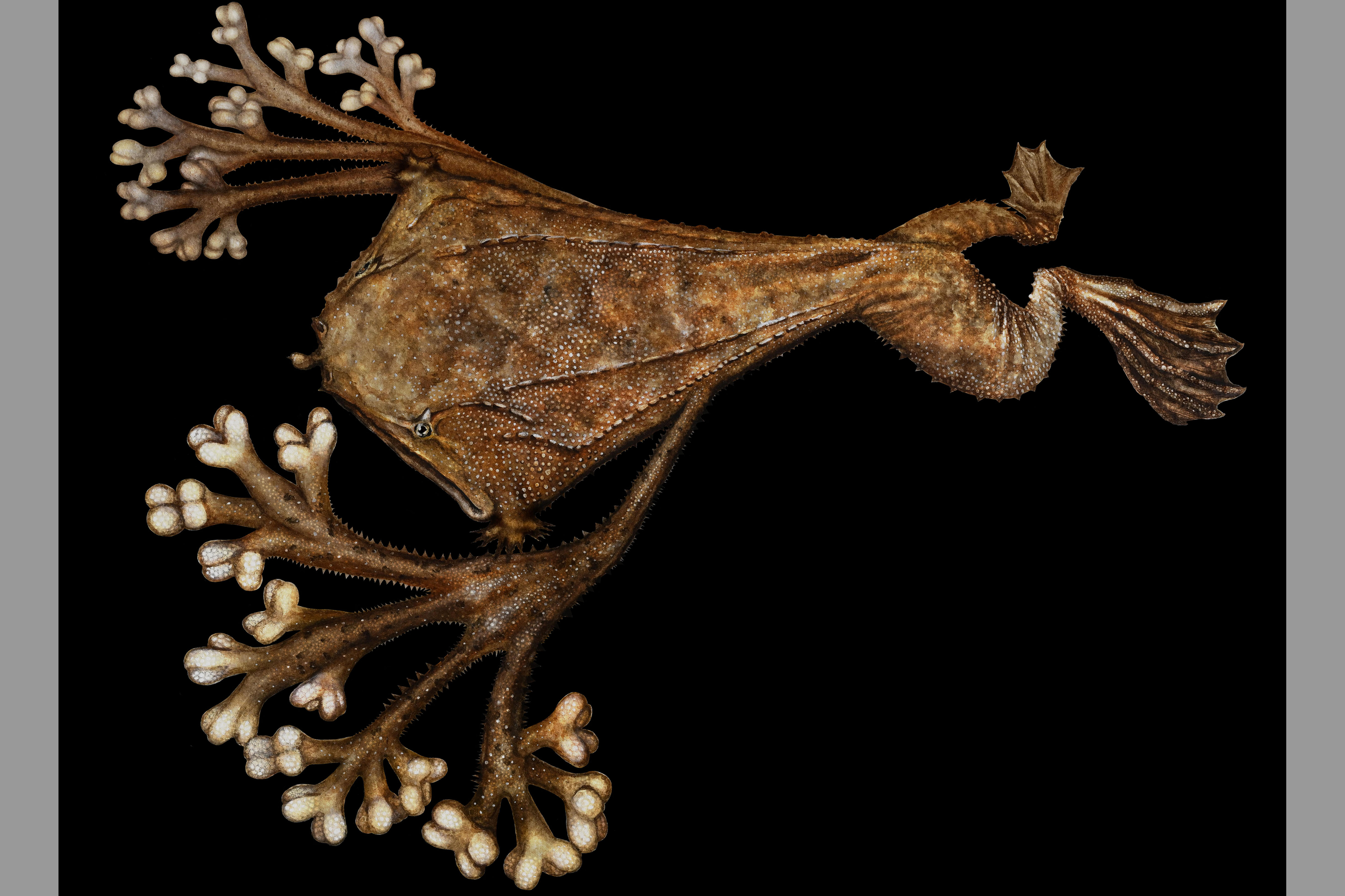
Surinam toads (Pipa pipa) are strange, flat frogs with minuscule eyes that live in the murky waters of the Amazon basin and carry their eggs on their back. The way they catch their prey is even weirder. They lie in wait with outstretched forelimbs until a small fish approaches, then snap open their jaws to create enough suction to draw the fish into their tongueless mouths and swallow it whole, all in a few hundredths of a second. But how do these frogs hunt in low visibility with poor vision?
A new study by UCLA biologists points to an answer that has been hiding in plain sight for two centuries: the frog's peculiar fingertips. In fact, they are commonly called star-fingered toads. The research, published in the Journal of Comparative Physiology A, has revealed a sensory system that processes signals gathered by neurons in unusual lobes on the frog's front fingers to identify the proximity and size of potential prey.
Because vision is not useful in their turbid environment, the frogs position their arms in front of and just to the sides of their mouth, fingers spread wide, to detect prey through the movement of water as a fish swims. Each finger ends not in a claw or pad but in a small cluster of softer lobes, an anatomical oddity noted by 19th-century naturalists but never satisfactorily explained. The new work shows that these lobes are dedicated touch organs, the amphibian equivalent to the fovea — a high-resolution patch of sensors at the center of the eye.
"I was reading an anatomy report that described how the tips of the fingers of Surinam toads split into four, and each of those four little lobes split again into four," said Duncan Leitch, a corresponding author and UCLA assistant professor of integrative biology and physiology. "It immediately seemed to me like these lobes might be somewhat analogous to antennae that the frogs extend so they can feel the space around them."
16 lobes per finger
Leitch and his co-authors looked at the anatomy of the finger surface area using a scanning electron microscope and identified a total of 128 mini-lobes, called lobules, per frog. The skin on each lobe was covered with nearly four times the density of dome-shaped bumps called papillae than on skin elsewhere on their fingers. Papillae, also found on the human tongue, are known to increase touch sensitivity.
They then touched the frog's hands with calibrated filaments to see how easily neurons from each skin area become excited by precisely known forces, and found that they became increasingly sensitive to touch, peaking at the ends of the fingers where thresholds fell into the same range as human fingertips. Next, the researchers studied the electrophysiology of nerves in the arms and found dense clusters of touch-receptive nerves on the fingertip lobules. Even though the lobules occupied only 8% of the total forelimb skin surface, they contained 60% of the touch-sensitive nerves serving the arm.
For the next phase of the study, the researchers observed high-speed video of the frogs hunting and consuming prey. The videos showed that when fish and other moving animals approached the frog's outstretched fingers within about half a centimeter, the frog could accurately and efficiently suck up prey before even touching it — even in the dark. This indicates that the frogs were using their fingers to detect the water's movement as the fish swam.
An exaggerated map in the brain
The results establish the fingertip lobes of the Surinam toad as specialized mechanoreceptive organs analogous to the tentacle-like appendages on the star-nosed mole's face. These types of specialized sensing organs are known as tactile fovea. This degree of magnification of important sensory surfaces has only been observed physiologically in several animals, including the bill of the platypus and the fingertips of primates — in a higher relay of the brain in the cortex, which is found only in mammals.
Frogs, however, don't have a cortex. The researchers instead found it in a part of the brain called the optic tectum, a midbrain area found in most vertebrates, where a region corresponding to just fingertip touch was greatly exaggerated.
The researchers determined how much space, if proportioned according to the entire body surface, this area should occupy. Even though the fingertips make up very little of the body's total surface area, the optic tectum region devoted to processing information from the fingertips was far out of proportion to their size.
Finding the same organizational logic in a frog — in a lineage that split from mammals more than 350 million years ago — suggests this magnification is a very old solution to a general problem: When an animal needs fine detail from a tiny patch of the body, evolution builds a fovea, whatever the body plan.
"It seems that mammals share some of these sensory processing properties with frogs, from whom they diverged evolutionarily a very long time ago," Leitch said. "This is probably a much more ancient and widespread kind of system than previously believed. People may have thought that these are mammal-specific traits, but certainly frogs, and I expect birds and other animals also would have specialized sensing systems if you really look for them."


