The liquid chromatography component of a mass spectrometer in the Vinogradova lab. (Credit: Lori Chertoff)
The body's organs are in constant communication. Fat tissue tells the liver when to store or release energy, the immune system signals localized inflammation, and thousands of proteins carry these messages to organs throughout the body. But while scientists have long known these conversations exist, they have struggled to identify exactly which cells are sending which messages.
Now researchers have found a way to listen in. By refining an existing protein-tagging technology called "proximity labeling", and applying it in genetically engineered mice, the team developed a platform that traces proteins back to their cells of origin and tracks them as they are manufactured and folded in the endoplasmic reticulum (ER). This allowed them to create the most detailed map to date of how fat and liver cells communicate, and how those networks are rewired during inflammation, fasting, and obesity. Upon cross-referencing their findings with data from the UK Biobank, the researchers linked 65 of these signals to human conditions, including type 2 diabetes and cardiovascular disease.
However, the study's most important contribution is not the discovery of any single protein messenger, the researchers note, but rather the creation of a powerful new framework for studying how organs communicate, opening the door to discovering new biological pathways and identifying signals that could serve as future biomarkers or therapeutic targets for any number of diseases.
"The platform is very broadly applicable," says Ekaterina V. Vinogradova, head of the Laboratory of Chemical Immunology and Proteomics. "It can be applied to different tissues and different genetic drivers-biologists in any field can use it to study cell-to-cell communication."
Combining expertise
A major obstacle to eavesdropping on organ communication is that scientists have only been able to meaningfully listen to one side of the conversation. The bloodstream is filled with protein messengers carrying instructions between tissues, and researchers can often detect the signals. But identifying exactly which cells sent each message has proved challenging.
Some researchers have tried measuring RNA inside cells as a proxy for the proteins that they produce; others have studied isolated cells in the lab or used chemical tagging methods to label proteins before they enter circulation. But RNA levels often fail to predict which proteins a cell ultimately secretes, labeling techniques often fail to detect less abundant signaling molecules, and cultured cells cannot fully replicate the physiology of living organisms. "A major limitation in the field has been how to discover these proteins in vivo," says Paul Cohen, head of the Weslie R. and William H. Janeway Laboratory of Molecular Metabolism.
The collaboration that would ultimately overcome that limitation began with shared frustration. Ken H. Loh, then a Rockefeller postdoc in the laboratory of Jeffrey M. Friedman, and currently an assistant professor at Yale, was interested in studying communication between adipocytes and nerves residing within fat tissue. He had genetically engineered a mouse model that promiscuously tags proteins as they transit through the endoplasmic reticulum. This had the potential to allow researchers to trace secreted proteins found in the bloodstream back to the specific cells that produced them by tagging those proteins as they passed through the endoplasmic reticulum, and Loh and Friedman shared it with Cohen and Vinogradova catalyzing the collaboration. But as each of the labs began working with the mice separately, they found that they were hitting the exact same technical roadblocks: they struggled to successfully enrich the tagged proteins and prevent rare signals from being lost in the mass spectrometry data. The mice were doing their job, but the challenge was extracting and detecting the tagged proteins with enough sensitivity to be useful. "We realized that, by joining forces, we could create a more impactful story together," Cohen says.
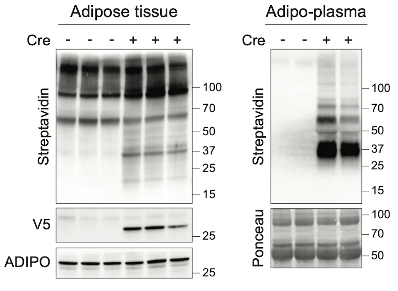
Figure 1C in the paper, showing western blots identifying proteins in adipose tissue and plasma. (Cohen lab)
Optimizing the platform
Combining their expertise, Loh and Cohen's group contributed physiological models of fasting, inflammation, and obesity, while Vinogradova's laboratory refined the tissue processing workflows, proteomics, and computational pipelines needed to isolate and identify tagged proteins. Together, the team improved how the platform recovered proteins from tissues and blood and reduced missing values in the mass spectrometry data. The result was a far more sensitive platform, capable of detecting low-abundance signaling molecules in the circulation that had escaped previous studies, including trace levels of the fat-derived hormone leptin.
"Without optimization, the platform identified only the most abundant proteins, which generally are the things people already know about," Cohen says. "It's only by refining the technique that we were able to get deeper into the unknown areas of biology. It's not just having the technology, but doing it really rigorously and well, that enables these kinds of findings."
The team then put the optimized platform to work in fat and liver cells, tracking how those tissues responded to fasting, inflammation, and obesity. To capture the progression of metabolic disease, the researchers compared the protein communication networks of visceral fat surrounding internal organs and subcutaneous fat beneath the skin during both early and advanced obesity, and also demonstrated the platform's broader applicability to immunology by successfully mapping protein production in B lymphocytes at baseline. They chose to study fat, liver, and immune cells because they needed a well-understood testing ground to validate their newly optimized platform. In addition, these choices reflected the interests of both laboratories: Cohen studies how organs, and particularly adipose tissues, communicate to regulate whole-body metabolism; Vinogradova uses chemical proteomic technologies to study immune protein function.
"Adipose tissue is a humoral hub in metabolic regulation, and its secretory repertoire clearly extends beyond known circulating factors such as leptin and adipsin," says Kaja Plucińska, a a postdoc in the Cohen Lab. "The tool we developed allows us to precisely trace and quantify novel, low‑abundance, potentially disease‑modifying blood‑borne adipokines and to characterize the secretome of less well‑studied endocrine fat cells, such as brown adipocytes, which we are very excited about."
In addition to cataloging proteins released into the bloodstream, the team also mapped proteins within the ER, where proteins are manufactured and folded, providing an unprecedented view of both the messages being sent and the machinery that produces them.
"People haven't been focusing on this, but it is important for both health and disease," Vinogradova says. "That's because it tells us not only what messages cells are sending, but also how the machinery that produces those messages changes under stress." The data revealed that the ER's protein folding machinery actively adapts to changing physiological conditions: fasting suppressed the production of many immune-related proteins, while severe inflammation triggered stress-response pathways that help cells cope with increased protein-folding demands.
The resulting atlas revealed distinct communication programs for each metabolic state studied and uncovered previously unknown factors, including γ-synuclein, a protein previously associated with the nervous system that emerged as a fat-derived messenger, declining during fasting and inflammation but rising sharply during advanced obesity, and MTR1L, an orphan receptor whose production spiked 30-fold in visceral fat during advanced obesity. They also discovered many proteins altered by obesity that had not previously been studied in relation to it. When the team compared their findings with data from more than 53,000 individuals in the UK Biobank, they found that 65 of the proteins that they had shown to be altered by metabolic stress were associated with human diseases, including type 2 diabetes, obesity, hypertension, coronary artery disease, heart attack, stroke, atrial fibrillation, and sepsis.
"On the side we worked on, which was proteins that are made by fat cells and regulated by obesity, we confirmed that the technique works," Cohen says. "But we also saw over 150 other proteins, many of which have never been studied in this context. We think that figuring out how those proteins regulate whole-body metabolism will be a really productive area, going forward."
"These TurboID experiments required a different approach to data analysis than our other mass spectrometry projects," says Charlotte Wayne, a postdoc in the Vinogradova lab. "But with the pipelines now established, we're excited to apply this strategy to additional cell types and disease settings."
Cohen plans to use it to explore what proteins brown fat secretes and uncover exactly how exercise improves whole-body health, while Vinogradova will extend the approach to other systems, particularly in immunology, where tracking cell-to-cell communication has proven particularly challenging. In his new lab, Loh has found that-like at Rockefeller-the mouse model has catalyzed new collaborations to study questions at the interface of ageing, obesity, and reproductive physiology.
"What excites me the most are the applications to study immune signaling and dysfunction," Vinogradova says. "But we're hoping that it opens the door to building an organ interactome across the entire spectrum of health and disease."