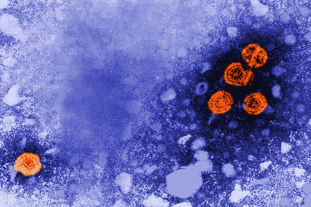
Transmission electron microscopic image showing hepatitis B virus virions in orange (Credit: CDC/Dr. Erskine Palmer)
Some 254 million people live with a chronic hepatitis B (HBV) infection that is often asymptomatic for decades, only to emerge in an advanced stage of disease that turns to fatal cirrhosis or liver cancer in nearly a million people every year.
The development of effective treatments for HBV infections has been stymied because we lack a good small animal model for studying the entire viral lifecycle, host response, and disease progression.
For decades, the assumption has been that the barrier to creating a workable mouse model is an inability for human HBV to gain a genetic foothold in the mouse due to its unique type of DNA. Now researchers from the lab of Rockefeller's Charles M. Rice have overturned this notion by discovering that the problem is something different altogether-likely a misstep in the entry process that renders the virus unable to deliver its DNA to the mouse cell nucleus. This work, recently published in PNAS, is funded in part by Rockefeller's Stavros Niarchos Foundation (SNF) Institute for Global Infectious Disease Research.
"This new understanding of the mechanisms behind human HBV's inability to infect mice provides a foundation for the development of fully HBV-susceptible mouse models," says Rice, head of the Laboratory of Virology and Infectious Disease. "Being able to model HBV's infection processes, host response, and disease progression could enable the design of new therapies for what is now a largely incurable disease."
Why HBV infections are chronic
HBV is a scientific conundrum: tiny, ancient, and unique, with an unusual replication process that generates a stable viral genetic template called covalently closed circular DNA (cccDNA), which is formed inside the host cell nucleus. Once acquired, these molecules are hard to get rid of-they're the reason for chronic HBV infection. They're also essential for viral gene expression and subsequent replication; without them, the virus is dead in the water.
Though various mouse models have existed going back some 30 years, none have the ability to support chronic HBV infection except humanized mice, which are engineered to carry human liver cells. One widely accepted theory in the field is that because mouse liver cells can't produce cccDNA, the virus can't replicate and spread from one cell to another.
However, Xupeng Hong, first author on the study and a postdoc in the Rice lab, found exceptions in the published literature showing that cccDNA was able to form in a mouse model-at least under certain experimental conditions-through a unique process in the HBV lifecycle called the intracellular amplification. This process recycles progeny HBV DNA back to the nucleus to form cccDNA, helping maintain cccDNA levels in infected cells and contributing to persistent infection. To Hong, that suggested the barrier to creating an HBV-infected mouse was not a settled matter but instead undetermined.
Moving closer to a model
To elucidate the true mechanism, Rice's group designed a method of creating a range of stable cell lines that supported HBV replication. This approach allowed them to sidestep the virus entry process in mouse liver cells and focused on post-entry steps and cccDNA formation via the intracellular amplification pathway. To their surprise, they discovered that all mouse cells tested were indeed capable of supporting cccDNA formation at levels comparable to that observed in human cells.
"This tells us that mouse liver cells can form the HBV's cccDNA, but to detect it, there has to be an abundance of them," Hong says. "My theory is that these have been in the cells all along but were unobserved due to low numbers."
Further experiments in which the HBV receptor was added to mouse liver cells helped narrow the likely location of the problem to a late step during the viral entry process. While Hong could not pinpoint the exact mechanism, the block occurs before nucleocapsid uncoating, a step in which the virus releases its DNA after entering cells.
When it comes to this nucleocapsid uncoating, timing and location are everything: If it happens too soon, the viral genome becomes visible to cellular sensing, and therefore becomes vulnerable to host cell antiviral defense; if it happens too late or in the wrong place, the virus may fail to establish cccDNA. So any change that precedes the uncoating may disrupt this process, leading to the virus's doom.
The lab's current research aims to ferret out exactly what this mechanism may be; identifying-and overcoming-it would hasten the creation of a new mouse model and allow for the observation of chronic hepatitis and cancer development. Ultimately, the goal is to figure out how to eliminate cccDNA from an infected person for good.
"A next-generation model would not only allow us to finally study the infection and host-virus interaction in detail, it would also provide a much-needed testing ground for new therapies," says Rice.


