Key Takeaways
- Targeted alpha therapy (TAT) is an emerging cancer treatment that uses alpha-emitting radioisotopes to target and destroy cancer cells without damaging nearby healthy cells.
- The radioisotope actinium-225 shows promise for TAT, but the medical scanners available today lack the resolution and detection efficiency needed to accurately image actinium-225 and locate tumors.
- A new technique called Time-of-Flight Cascade Gamma Imaging (TOF-CGI) could allow existing scanners to image TAT accurately and enable its widespread adoption.
A research team led by Lawrence Berkeley National Laboratory (Berkeley Lab) has developed a new imaging technique that could allow modern medical scanners to image targeted alpha therapy (TAT), an emerging cancer therapy for the diagnosis and treatment of cancer. The advance could enable the widespread adoption of this powerful new cancer therapy.
"TOF-CGI brings us a step closer to helping clinicians diagnose and treat cancer in a way that wasn't possible before." - Javier Caravaca, staff scientist, Nuclear Science Division
Targeted cancer therapies use specially engineered radioisotopes that target and kill cancer cells. One of the most promising of these new approaches uses alpha-emitting radioisotopes such as actinium-225. Clinicians still lack a reliable way to image actinium-225 because the medical scanners available today lack the resolution and detection efficiency needed to detect the activity of alpha-emitting radioisotopes in the body. But now a new technique called TOF-CGI (time-of-flight cascade gamma-ray imaging) could close that gap. The work was recently published in the Journal of Nuclear Medicine.
"TOF-CGI brings us a step closer to helping clinicians diagnose and treat cancer in a way that wasn't possible before," said Javier Caravaca, a staff scientist in Berkeley Lab's Nuclear Science Division and principal investigator of the new study.
Advancing TAT imaging with TOF-CGI
Targeted radioisotopes are engineered with targeting molecules to form a radioactive compound. The targeting molecule guides this compound to a cancer cell, where it binds. Actinium-225 is a leading candidate for targeted alpha therapy because of its exceptional potency, and its half-life of around 10 days is ideal for localized treatment with fewer side effects. Actinium-225 emits four alpha particles as it decays, splintering a cancer cell's DNA into fragments without harming nearby healthy cells. This prevents the cancer cell from regenerating and slows the progression of disease.
Actinium-225 could also be a powerful imaging agent. As the radioisotope decays, its daughter products release gamma photon pairs; the idea is to use these signals to locate tumors while tracking how they respond to treatment over time. Advanced algorithms allow medical scanners to reassemble such data points into 3D images of a tumor.
"Traditionally, there have been only two ways to image cancer: SPECT (Single-Photon Emission Computed Tomography) or PET (Positron Emission Tomography). Although these techniques have led to big advances in cancer imaging, neither have so far demonstrated the demanding requirements to image targeted alpha therapy accurately. Our new study is the first to show that there could be a third way to image cancer with the alpha therapy radioisotope actinium-225," Caravaca said.
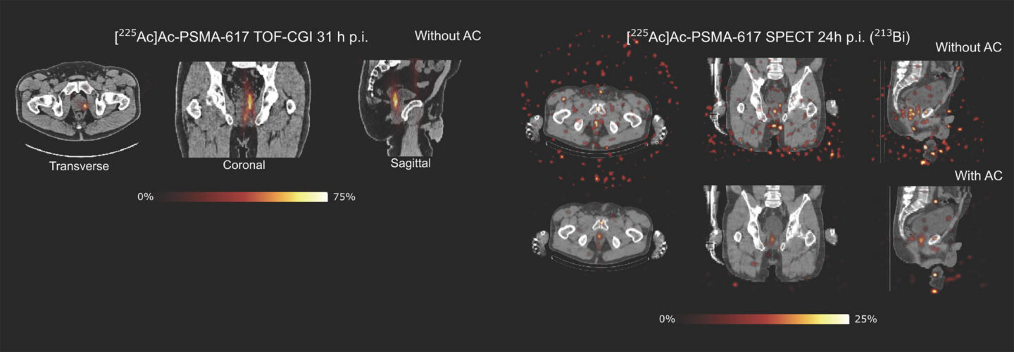
SPECT scanners are commonly used in hospitals, but they lack the required sensitivity to accurately generate images from the detected gamma rays in targeted alpha therapy. SPECT uses thick, heavy metal shields (collimators) to detect single gamma rays - but this only works well when the flux of gamma rays is large. And because targeted alpha therapy is so powerful, only small doses of the radioisotope - about 100 times less than a conventional radiotracer - is administered to patients. Altogether, these constraints add up to low gamma-ray sensitivity and noisy images.
On the other hand, PET promises higher gamma-ray sensitivity and image quality because it doesn't use collimators. PET works by picking up "positron annihilation" events - When a positron collides with an electron, it emits two gamma-ray photons back-to-back. The PET algorithm quickly combines tens of thousands of these data points to generate an image of the radioisotope accumulation in the body, such as in a tumor.
Researchers have long assumed that PET scanners wouldn't work with actinium-225 because the radioisotope emits alpha particles, not positrons. "But one day we wondered, what if we could exploit actinium-225's gamma-ray pairs to demonstrate targeted alpha therapy in a PET scanner?"
So Caravaca wrote a new algorithm from scratch - TOF-CGI is an image reconstruction algorithm that uses the detected gamma ray pairs to pinpoint where actinium-225 decayed, and reconstruct that data into a 3D image of actinium-225 in the body. TAT emits thousands of signals as the radionuclide decays, so leveraging the GPU computation capabilities at the National Energy Research Scientific Computing Center (NERSC) was essential to significantly accelerate the image-generation process during the development and testing of this technique.
Caravaca and co-authors at UC San Francisco then conducted a pilot study with a patient who was previously diagnosed with prostate cancer through standard PET scans. A validation test confirmed that TOF-CGI in a PET scanner identified the same prostate tumors that were detected by the initial PET diagnostic scans. Subsequent experiments showed that TOF-CGI in PET detected more actinium-225 decays in the patient than SPECT, resulting in clearer images. The results represent the first TOF-CGI imaging of a human subject.
"This is an exciting first step. Clinicians want to know whether a radioisotope is doing its job. Did it stop the cancer cells from spreading? Or did new tumors emerge? Targeted alpha therapy imaging could help us answer those questions and more, but we still need an imaging modality to match. We think that our TOF-CGI technique could one day help us get there," Caravaca said.
In future studies, Caravaca and team hope to expand the new technique to other radioisotopes and test its efficiency with a full-body PET scanner.
Researchers from Berkeley Lab, UC San Francisco, and Siemens Medical Solutions USA Inc. contributed to the study.
TOF-CGI was recently awarded a 2026 R&D 100 Award and is available for licensing. Contact