
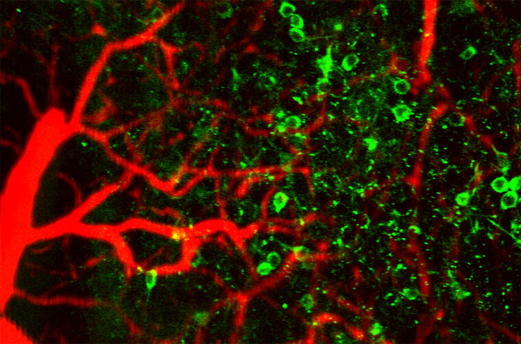
The brain is a black box of densely packed neurons, blood vessels and immune cells, all locked away in a sealed skull. WashU's Song Hu has dedicated his career to unlocking that puzzle box by developing new ways to "see" inside. His team's newest innovation could give researchers a new view of how brain cells and blood vessels work together and what happens when the coupling between the two breaks down.

Hu, a professor of biomedical engineering at Washington University in St. Louis, and his team at the McKelvey School of Engineering have developed a new way to integrate two powerful techniques: two-photon microscopy and photoacoustic microscopy (TPM-PAM). This will allow researchers to see in real time how the brain vasculature delivers oxygen to neurons. The new system could have profound implications for studying neurodegenerative disease, stroke and other diseases connected to neurovascular coupling.
"Using this technology gives us a better understanding of these disease mechanisms," Hu said.
Hu and his team shared their results in the journal Nature Communications, including how they combined the two different forms of microscopy into one system and successfully recorded, for the first time, single-neuron calcium activity alongside oxygen release from individual red blood cells in awake mice.
Neuronal activity and oxygen delivery in action
Two-photon microscopy is widely used to image neuronal activity in a brain that is lit up by fluorescent probes.
The other technique, photoacoustic microscopy, uses light-generated sound waves to record blood flow and oxygenation. Brain function is intricately linked to its vasculature. Thinking itself consumes energy, but without the view of oxygen delivery that PAM can provide, it's very difficult to understand any details about how the fuel pipeline works in relation to brain activity.
"That's why it's important to image both of them, to understand how neuron activity consumes oxygen and how the vasculature changes its dynamics to meet the demand in real time," Hu said.
With this new technique, Hu and his team were able to bring the two complementary views together at cellular resolution for the first time.
"Combining the two for simultaneous imaging of neuronal activity and oxygen delivery, at single-cell level, has never been demonstrated before," Hu added.
To demonstrate the platform, the team imaged the brains of mice while stimulating their whiskers, tracking neuronal calcium activity alongside changes in blood oxygen delivery. The researchers also used tightly focused laser pulses to block a single capillary or stimulate a single neuron, then followed the resulting changes in nearby neurons and red blood cells. Together, these experiments show how TPM-PAM can combine precise, cell-scale interventions with simultaneous measurements of neuronal activity and oxygen delivery, allowing researchers to move beyond observing correlations and begin probing cause and effect.
Although demonstrated so far only in animal models, the platform could help reveal how brain cells and microvessels coordinate energy use and how that relationship is disrupted in stroke and neurodegenerative disease.
This platform also could inform clinical imaging. Functional MRI does not directly record neurons; it infers changes in brain activity from blood oxygenation and flow. A clearer cellular-scale picture of neurovascular coupling could help researchers interpret these indirect signals much more accurately.
"If you can better understand neurovascular coupling, you will make this inference process more accurate," Hu said.
Making room for light and sound
The technical challenge was solved with help from engineering colleagues at Northwestern University.
Light and sound waves don't usually play well together. "If you want to integrate the two, you have to make the two forms of energy share the same space, and this is not easy," Hu said.
Conventional PAM systems use ultrasound detectors that block the optical path or require optical lenses that sacrifice two-photon resolution and light collection. The team's solution is an optically transparent acoustic sensor built from a polymer micro-ring resonator on a piece of glass. When ultrasound impinges on the resonator, it slightly deforms the ring and shifts its optical resonance, converting the acoustic signal into a measurable change in light. At the same time, the transparent sensor allows TPM's excitation and fluorescence light to pass through with minimal interference.
"It's not compromising any of the two imaging technologies," Hu said.
Han J, Lee Y, Feng Z, et al. Integrated two-photon and photoacoustic microscopy for single-cell neurometabolic imaging. Nature Communications. https://doi.org/10.1038/s41467-026-75603-7
This work was supported by the National Institutes of Health (R01 NS120481, AG079503, and NS125677 to S.H.; P41 GM135018 to H.F.Z. and C.S.) and the Washington University Imaging Sciences Pathway Fellowship (to J.H.).


