
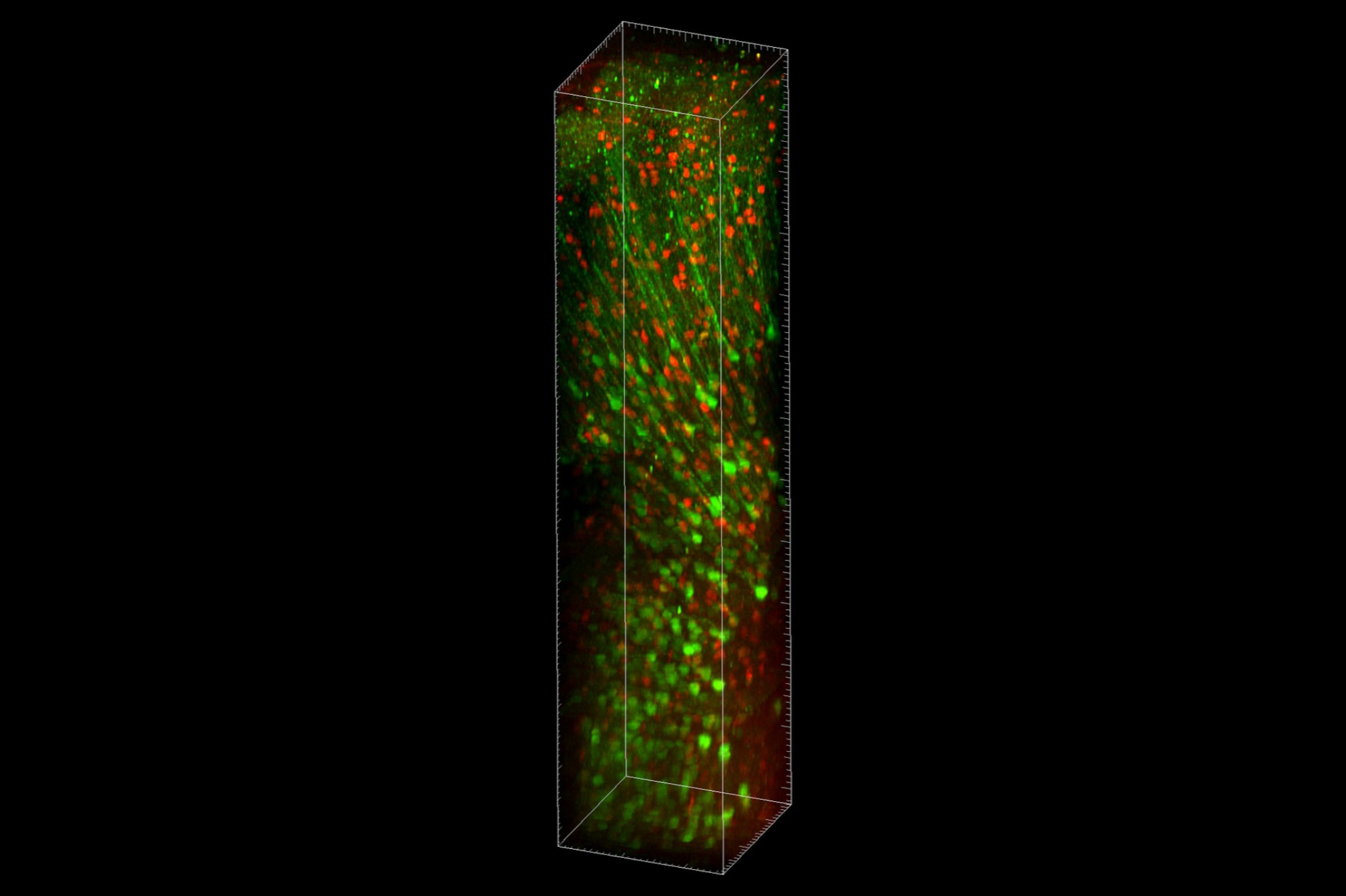
A volumetric rendering of stacked images taken with the Vaziri lab's new platform TrUDI. (Credit: Vaziri lab)
As light travels deeper into the brain, it scatters. Three-photon microscopy, the current state of the art, reduces the resulting background fluorescence and can, in principle, image much deeper than earlier techniques-but to do that, it uses average laser power limits that cause overheating and damage living tissue.
Instead of accepting that tradeoff, a Rockefeller team has now redesigned the entire imaging system from the ground up. In a paper published in Nature Methods, the team describes an innovative microscopy platform called Three-photon Ultra-Deep Imaging (TrUDI), which pushes three-photon imaging close to its theoretical limits, in part by reengineering the rate and energy of laser pulses, how they are delivered and scanned at the sample-all within the safe limits of sample power exposure. The result is a roughly 250-fold increase in imaging efficiency, allowing researchers to image through the entire mouse cortex and record neural activity in deep brain regions, such as the entire depth range of hippocampus and striatum, without surgically removing the overlying tissue.
"Often in technology, people stick with a certain laser or parameter for historical reasons, and everyone just accepts those limitations," says Alipasha Vaziri, head of the Laboratory of Neurotechnology and Biophysics. "We wanted to take a clean slate approach. By not taking anything for granted and asking how a system fully optimized for maximizing the depth reach of three-photon imaging would look like from scratch, we found that what people previously thought to be limiting factors could be overcome."
A clean slate
One of neuroscience's greatest challenges is understanding how disparate groups of neurons deep in the brain work together to produce behavior. Although modern microscopes can record the activity of individual neurons near the brain's surface in living animals, imaging broad swaths of neurons deep inside the brain has remained difficult, because brain tissue scatters light. As light travels deeper, it becomes impossible to distinguish neurons from background fluorescence.
For years, researchers have pushed this limit using increasingly sophisticated forms of fluorescence microscopy, in which engineered molecules emit light when illuminated by a laser. Three-photon microscopy, among the greatest achievements in this space, can in theory image much deeper than other methods, including the more widely used two-photon microscopy, because it generates less background fluorescence. But reaching those depths with three-photon microscopy faces practical limits.
"That higher demand for pulse energy in three-photon excitation ultimately sets a limitation on how much laser power you can put into the brain at typically used laser repetition rates before damaging it," Vaziri says. "You are limited by heat, and at the same time, the number of photons reaching the target drops exponentially due to scattering, causing the fluorescence signal to become indistinguishable from the background."
Three-photon microscopy was designed to extend the ~0.8 mm depth reach of the more widely used two-photon microscopy, but it has been falling short of its potential. "In any multi-photon excitation scheme, there is a depth limit at which the background and the signal become equal. For three-photon excitation in the mouse brain, theoretically that depth limit should be on the order of 2.3 millimeters," Vaziri says. "But the maximum depth at which functional recording has been previously achieved was about 1.2 millimeters."
Principled optimization
Vaziri resolved to redesign the imaging system from the ground up. His team began by using computer models to determine the theoretical limits of deep-brain imaging and identify how a microscope would need to be designed to approach them. Guided by those calculations, they went back to the drawing board and optimized every aspect of three-photon microscopy, most notably by delivering fewer but much more energetic laser pulses while maintaining the same average laser power, and by redesigning how the microscope focuses and collects light. They even developed an excitation scheme that identifies neurons and activates the laser only when passing over those cells, reducing laser exposure by up to 80 percent during long sessions.
"The way we did this was through a series of very systematic, principled, and ground-up optimizations," Vaziri says. "We tried to not make any ad hoc assumptions about the choices of the parameters of the microscope or the laser, having them all being informed by the ultimate purpose that they are serving, which is maximizing depth reach while faithfully resolving transient activity of individual neurons within safe laser exposure limits of the sample."
Together, these optimizations added up. The final product, the TrUDI platform, boasts a roughly 250-fold increase in fluorescence generation efficiency without relying on increased laser power that could heat and damage brain tissue. "When you look at individual parameters, an optical tweak might only give you an improvement of a factor of four or five, which is easy to overlook when you are up against an exponentially hard problem," he says. "But if you have enough of these optimizations and each of these improvements is multiplicative, then you suddenly have something that is 200-fold better."
Using TrUDI, the team then validated the new system in mice, imaging the activity of neuronal populations across cortical depths into deep structures of the hippocampus and the striatum, reaching tissue depths of up to 2.1 millimeters in the intact mammalian brain-far beyond previous limits, which had stalled around 1.2 millimeters. For the first time, the team captured activity across multiple interconnected regions of the hippocampus and deep within the striatum without surgically removing the overlying cortex.
Analyses of brain tissue designed to detect heat-related injury and other signs of damage found no evidence of inflammation, neuron loss, or DNA damage under the imaging conditions used in the study, confirming that TrUDI can safely reach depths previously considered impractical for optical imaging.
As a result, TrUDI also provides a window into brain circuits that have, until now, been largely unreachable. "There are regions of the striatum that are inaccessible, and there are still a number of deep brain nuclei which are much deeper that still require invasive approaches," Vaziri says. "But our work brings a series of brain regions that have been previously accessible to imaging only through invasive means within reach, without that kind of invasiveness."


