Targeted activation of specific nerve cells using pulses of light alleviates motor impairments in an animal model of Huntington's disease and restores healthy neuronal activity. These findings have now been published in the journal Nature.
Huntington's disease is a hereditary disorder caused by a mutation in a single gene, which remains incurable to this day. Patients primarily suffer from a severe loss of motor control, characterized by involuntary, sudden, and unpredictable movements of the arms, legs, face, and trunk. In addition, psychiatric symptoms such as depression, irritability, or aggression are common, as is a progressive decline in cognitive abilities that can ultimately lead to dementia.
According to Germany's AOK health insurance association, around 10,000 people in Germany are affected by the disease, with several hundred new cases diagnosed each year. Life expectancy of the patients is reduced, and the most common cause of death is swallowing impairment and its resulting complications.
Researchers Identify an Important Neuronal Cell Type
In a new study, an international research team has now identified a neuronal population that plays a key role in these motor disturbances. Furthermore, the scientists succeeded in ameliorating the movement defects. This discovery marks a strategic turning point: while research for decades has focused primarily on neuronal cell death, the emphasis is now shifting toward dysfunctional neuronal activity.
The study was led by Professor Takaki Komiyama from the University of California San Diego (UC San Diego) and Professor Irina Dudanova, who holds the Chair of Anatomy and Cell Biology I at Julius-Maximilians-Universität Würzburg (JMU) since April 2026. The study's first author is Dr. Sonja Blumenstock, currently a postdoctoral researcher at UC San Diego.
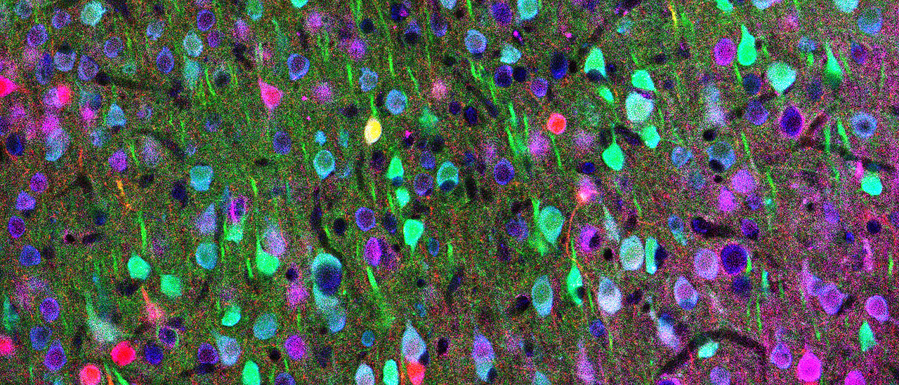
The team focused on cortical interneurons, which have so far received little attention in Huntington's disease research, as well as on so-called corticostriatal projection neurons (CStr) - nerve cells that function as a "data highway" connecting the brain's motor planning center, the cerebral cortex, with the striatum, the region responsible for executing movement.
"This system is like an orchestra: in a healthy brain, all instruments play in perfect synchrony. In Huntington's disease, however, the orchestra loses its rhythm," explains Irina Dudanova. The cortex is among the earliest sites affected by the disease. Here, the first dysfunctional signals appear long before the cells start dying.
Imbalance Among Inhibitory Neurons
The research team identified a fundamental imbalance among inhibitory nerve cells - known as interneurons - that was linked to the movement defects. This dysfunction occurs not only during complex tasks, but during all kinds of movement, whether structured locomotion training, spontaneous everyday movements such as scratching, or even resting states.
"There are different types of nerve cells in the brain that work together like a chain to control movement," Sonja Blumenstock explains:
- CStr output neurons: These cells send the actual "Move!" command to the body.
- SST cells: These act as "brakes" and suppress movement-related signals.
- VIP cells: These function as "supervisors." Their task is to switch off other inhibitory cells and thereby, metaphorically speaking, release the brake.
"In the healthy brain, VIP cells trigger a cascade that ultimately enables the CStr output neurons to initiate movement," says Irina Dudanova. In Huntington's disease, this chain is disrupted. As a consequence:
- VIP cells become almost completely silent.
- SST cells become overactive because control by VIP cells is missing.
- CStr output neurons are suppressed so strongly by the overactive SST cells that communication with the action execution center is effectively blocked.
Light-Based Therapy Approach Corrects Dysfunction
To interrupt this vicious cycle, the team used a specialized technique known as optogenetics. In this method, cells are genetically modified to become light-sensitive and can then be controlled with high precision. In the study, for example, the researchers selectively stimulated VIP cells using red light.
The stimulation resulted in clear benefits: mice receiving light stimulation showed significantly improved motor function. Their gait became more regular, and the disease-typical dragging of the hindlimbs was markedly reduced. Particularly striking was the observation that the activity of the corticostriatal projection neurons returned to the level seen in healthy control animals.
"We were surprised to see how sustainably the light pulses stabilized the network. Our results demonstrate that mechanisms of learning and plasticity can be reactivated even in a diseased brain," says Sonja Blumenstock. Indeed, the beneficial effects persisted when the light source was switched off.
"Our study shows that precise intervention into the brain circuitry can lead to significant behavioral improvements. If we know which cells to target, we can re-tune the abnormal activity patterns. This gives hope for future therapies," adds Irina Dudanova.
"This work shows that correcting specific imbalances in brain circuits can restore function, even in a complex neurodegenerative condition, and highlights the potential of targeting defined cell types to promote recovery," says Takaki Komiyama.
At the same time, Irina Dudanova stresses that clinical application is still a long way off. Considerable further research will be required before findings from animal experiments can be translated into therapies for humans.
Original Publication
Restoring cortical disinhibition improves Huntington's disease phenotypes. Sonja Blumenstock, David Arakelyan, Nicholas del Grosso, Sonja Schneider, Yufeng Shao, Enida Gjoni, Rüdiger Klein, Irina Dudanova* and Takaki Komiyama*. Nature, DOI: 10.1038/s41586-026-10671-9


