Fibre laser microscopy for use in clinical applications
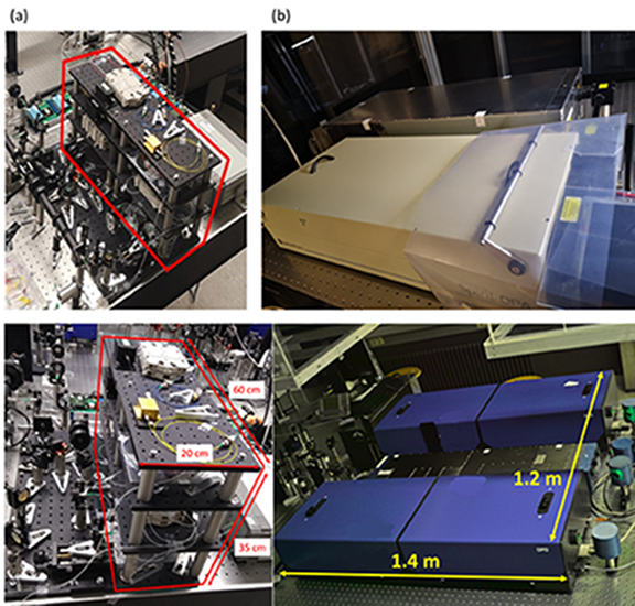
(a) Compact fibre laser sources for CRS imaging (in red box).
(b) The commercial laser sources for CRS imaging.
A research group led by Professor Kenneth K.Y. Wong of the Department of Electrical and Electronic Engineering at the University of Hong Kong (HKU), in collaboration with Bielefeld University in Germany, has developed a compact fibre laser microscope that brought breakthroughs to analysing molecules in cells and clinical applications.
The newly innovated microscope generates far less noise than customary designs, and the compactness and stability make it suitable for use in operating rooms in hospitals. The innovation was presented in the journal 'Light: Science and Applications,' published by Springer Nature.
When investigating how tumors grow, or how pharmaceuticals affect different types of cells, researchers have to understand how molecules within a cell react – and interact. This is possible with modern fluorescence microscopy. However, molecules in cell specimens had to be labelled with fluorescent substances to make them visible, and this can distort the very behavior of the molecules. Also the staining with fluorescent markers is generally unsuitable for in vivo tissues.
Label-free microscopic imaging has always been a hot topic in biomedical research. The newly invented laser microscope does not require fluorescent markers to obtain a clear image of cell molecules. Instead, cell molecules with different level of characteristics are uniquely presented via a Raman Imaging System.
Professor Kenneth Wong, who led the research said: "We use fiber laser as the light source of the optical microscope to replace the traditional solid-state laser, which is a brand new concept. Traditionally, the laser needs to be amplified in a free space of several meters, so the instrument is very big. With fiber lasers, light is amplified and transmitted through glass fibers, and the instrument design becomes light and compact. The volume is only one-eighth to one-tenth of the traditional solid-state laser instrument. Due to the size of the instrument, there is a limitation on where it can be used currently, but this will no longer pose a problem in future."
Professor Wong explained: "Fiber lasers were previously not favorable for microscopes because they were less powerful and very noisy compared to solid state lasers. To obtain molecule-specific imaging with their microscope, the team used two synchronized optical resonators (laser cavities), both with short picosecond pulses – one picosecond being one thousand billionth of a second."
"One challenge here was to control the lasers so that both beams with different wavelengths are synchronized, and hit the specimen at exactly the same time and position," said Professor Thomas Huser in Germany, a biophysicist at Bielefeld University.
Professor Huser believes that the new microscope is likely to be used in clinical applications in the coming years. Preliminary studies in cooperation with the Evangelisches Klinikum Bielefeld Hospital in Germany are already underway to use the microscope to analyze liver tissue samples."
"Our project partners are amazed by what this microscope can do." said Professor Huser. "Label-free microscopy can be used, for instance, to investigate how various new types of cells develop from stem cells. It also allows for a tumor to be demarcated from normal tissue without staining. Furthermore, we can ascertain how pharmaceutical compounds react with molecules in the muscle tissue cells of the heart and liver, as well as other cells."
Professor Wong believes the new technology can be applied in many biomedical applications, such as the endoscopy of the intestines and digestive system, etc., to detect early tumors and lesions.
"Using fiber laser, the image clarity can be 100 times higher than that of traditional endoscopes. It can penetrate the surface of organs and reflect the condition of deeper tissues. The light source uses harmless infrared visible light and will not affect the human body. In the long run, since it is portable, unmarked and harmless, it can be clinically used in surgical operations, such as immediate pathological detection, to mark tumor borders during an operation, or to accurately mark different parts for precise cuts during brain surgeries."
Lead author of the study Dr Cihang (Sherry) Kong said: "The prototype of the microscope will now serve as the basis from which to build portable devices. Because the molecules do not first have to be labelled, the specimen does not take a long time to prepare compared with using other microscopes, and the labelling-induced toxicity can be avoided. "Dr Kong is a former PhD student in Professor Wong's group, and currently a post-doctoral researcher of Professor Thomas Huser.
Both the University of Hong Kong and Bielefeld University have pioneering research in the biomedical and health technology fields, while HKU's Engineering Faculty is especially focused in the research of imaging technologies. This research was supported by the Germany/Hong Kong Joint Research Scheme sponsored by the Research Grants Council of Hong Kong and the Germany Academic Exchange Service of Germany, the European Union's Horizon 2020 research and innovation program under the Marie Sklodowska-Curie Grant, project "DeLIVER", the Research Grants Council of the Hong Kong Special Administrative Region, China, the National Natural Science Foundation of China, and the Innovation and Technology Fund.
Details of the paper:
High-contrast, fast chemical imaging by coherent Raman scattering using a self-synchronized two-colour fibre laser
Cihang Kong, Christian Pilger, Henning Hachmeister, Xiaoming Wei, Tom H. Cheung, Cora S. W. Lai, Nikki P. Lee, Kevin. K. Tsia, Kenneth K. Y. Wong and Thomas Huser
Light: Science & Applications volume 9, Article number: 25 (2020)


